| basic morphology of bdelloids: the mastax |
| The mastax is the organ in the digestive system of all bdelloid rotifers which transports the diet from the mouth to the stomach. Aside from muscles, nerve cells and digestive glands this organ also has some chitinous hard parts called trophi. (By analogy to the jaws and teeth of vertebrates one might think of the trophi as "grinding machine", but a closer look at the ingested diet shows that the trophi are not able to grind for example algal cells of fungal spores) |
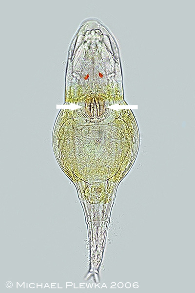 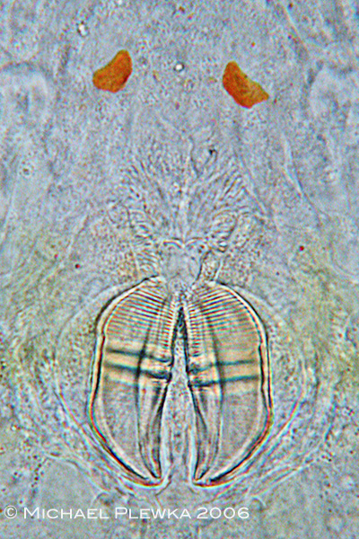 |
| The trophi are best visible in specimens which are compressed by the coverslide, either by mechanichal pressure or by removing the water under the coverslide. Left image: typical habitus of a compressed bdelloid rotifer (Philodina citrina) showing the trophi. The right image (crop of left image) shows more clearly that the trophi are typically connected on one side (here: on the anterior side) even when compressed. |
| |
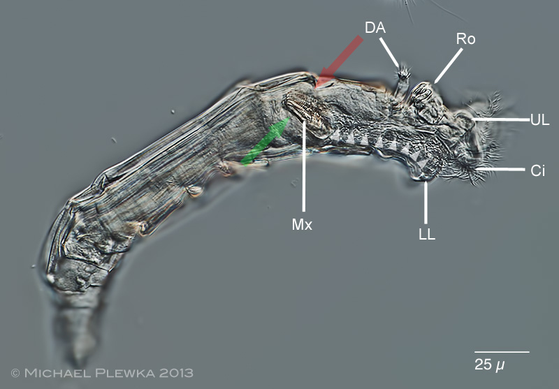 |
| This lateral view of a bdelloid rotifer (Macrotrachela musculosa) shows the orientation of the trophi in the mastax (Mx) in a living specimen while whirling. In contrast to the above image of P. citrina this image here shows the natural orientation of the trophi: the plane of the unci is typically in an angle of approx. 30-45 degrees relative to the longitudinal axis of the rotifer. The trophi can be looked upon from the frontal side ("cephalic view" (red arrow), see below) and from backwards ("caudal view", green arrow). DA: dorsal antenna; Ro: rostrum; UL: upper lip; LL: lower lip; Ci: cilia of the trochal discs |
| |
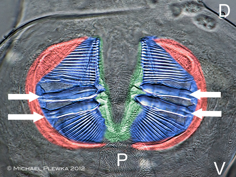 |
Artificially colored image of a compressed specimen of the very common rotifer Rotaria macrura. While in the larger parent taxon: Eurotifera there are many different types of trophi in the bdelloid rotifers only have a single type of trophi, which is called ramate. The image shows the ramate trophi in cephalic view (view from the anterior part); so the pharynx (P) yields the mastax with the trophi from the ventral side (the ventral side (V) is bottom, the dorsal side (D) is top). The colors characterise the different parts of the trophi. Red: manubria; blue: unci; green: rami.
The comb-like strucure of the unci is composed of chitinous teeth. The 4 arrows point to the major teeth, the number of which is a species-specific trait. The number of major teeth is therefore expressed as the so-called dental formula (DF) which is 2 / 2 for this specimen/ species. |
| |
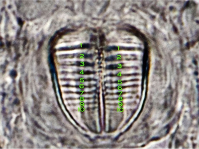 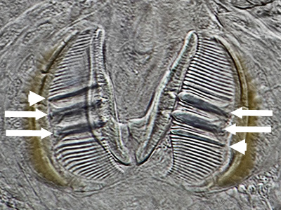 |
| Two more examples. Right image: Scepanotrocha semitecta with dental formula 10 / 10. Although the major teeth get less visible from top to bottom in this image they can be distinguished from the minor teeth because the latter ones are below the resolution of this microscope lens in this image. Right image: aside from the major teeth (marked by arrows) in the trophi of some species there are teeth in the size between major and minor teeth (marked by arrowheads). This is expressed in the dental formula which is: 1+2 / 2+1 in the case of the species Macrotrachela magna presented here. |
| |
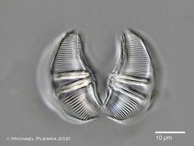 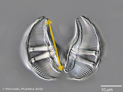 |
| For na undisturbed view on the trophi the maceration by NaOCl is mandatory. After maceration, depending of the focal plane, there is the aspect as viewed from the frontal side ("cephalic view"; left image) or the aspect from behind ("caudal view"; right image). Also by maceration it becomes evident that the major teeth (here: two in each uncus of Rotaria neptunia) consist of several thinner fibers which are close together. In caudal view (right) the rami are in focus. The size of the ramate trophi in bdelloids is species-specific, therefore the ramus length (marked in this image by yellow double arrow) is a quantitative trait that can help to characterize and separate species within genera). |
|
| Here is a general chart of the dental formulas of the trophi |
| Here is a list of other trophi types in rotifers |
| |
| |
| |
|
|
|
| |
| |